The Hidden Price of Competitive Gaming
Read more
7 months ago

|
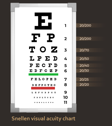
Eye check-up is normally carried out at the clinic/ office, using different equipment which helps to magnify the eye structure. It always starts with asking the patients what is the issue of concern, and associated symptoms which may be relevant. This is followed by visual acuity testing using internationally standardized chart. This step is very important in determining the course of further examination and is normally under five minutes. |
 |
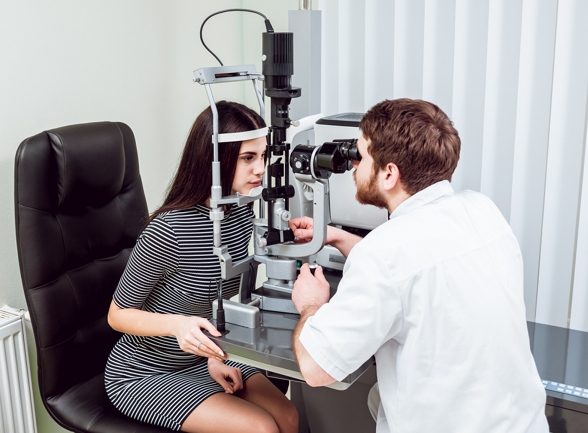
Patients are then asked to sit at the slit lamp biomicroscope for further examination. Patients need to be able to sit down and put their face over the slit lamp (photo). In particular, the Ophthalmologist will look at the coating of the eye, the cornea, and lens for any abnormalities which may contribute to the symptoms. This may take approximately 5- 10 minutes.
The next step is dilated eye examination to look at the retina, optic nerve, retinal blood vessels and other supporting structures including the vitreous. Pupil dilatation typically takes about 15 minutes, and patients are usually asked to wait- or sent for ancillary tests by the attending ophthalmologists. The doctor will use a magnifying lens to do this part of the eye examination.
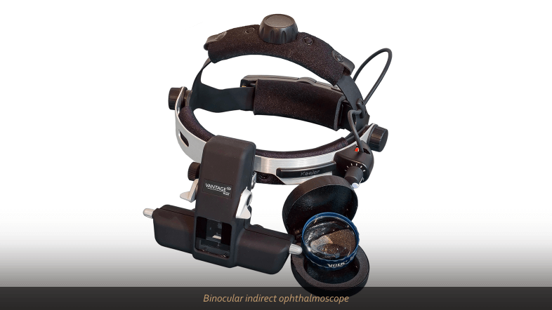
Children or patients with special needs may require longer time for examination. They may or may not be able to sit at the slit lamp, hence other equipment may be utilized such as hand-held slit lamp, and/ or binocular ophthalmoscope (photo).
Refraction or “checking the eye power” is also very commonly performed at the clinic by trained optometrists. This examination determines what is the patients’ best corrected vision and is important in determining the course of management and improving patients’ vision by glasses or contact lens prescription.
Intraocular pressure check is also done using contact or non- contact method to ensure that it is within normal limits. In cases of suspected angle-closure glaucoma, a special contact lens will be used to view the irido-corneal angle and assess the severity of angle closure. Other non-routine examinations are extraocular eye movement to check for normal alignment of both eyes, or to detect any double vision, and visual field examination.
 Common ancillary tests at eye clinic/ office
Some centers are equipped with anterior (front) segment photography which may be utilized to photograph any significant findings, and aid in the explanation of a patient’s condition. This also helps to document the disease progress or improvement. Similarly, posterior (back) segment photography serves the same function.
This test is done on a periodic basis to document any changes in the visual field and is normally used for patients with glaucoma or suspected/ confirmed neurological conditions. In glaucoma patients, this test is a must, to monitor disease progression and efficacy of treatment.
OCT of the macula provides a cross-section image of the retina which is very useful in determining cause of visual problems in diabetic retinopathy (in diabetes mellitus), age-related macular degeneration, or severely myopic (very short-sighted) eyes, to name a few. Another form of OCT imaged the thickness of the optic nerve and is important to look at the health of the nerve fiber layer.
This test is performed for patients who have cataract and want to proceed with cataract surgery. Ocular biometry is necessary for the calculation of intraocular lens, which will be implanted in patients’ eye during the cataract procedure. The doctor may request repeated measurement to ensure accuracy of lens calculation.
This is a specialized test, usually performed for patients with suspected macula-related visual problems, and supplements the findings from OCT. A specialized dye is injected intravenously and a series of retinal photography is taken. This test takes 20 minutes and the results can be determined on the same visit. Patients need to inform of any allergy to food or medication prior to the dye injection.
Dr Umi Kalthum binti Md Noh
Sunway Medical Centre
Jalan Lagoon Selatan, Bandar Sunway
47500 Selangor
DL: +603 7491 1080
LASIK enquiry: +6019 229 8672